Mostrando entradas con la etiqueta anatomy. Mostrar todas las entradas

domingo, 14 de febrero de 2016

Respiratory System.

Respiratory System.
The cells of our body consume oxygen in order to obtain energy. In this process, called oxidation, the cells burn glucose using oxygen, releasing carbon dioxide, and obtaining the necessary energy to carry out several metabolic processes. The respiratory system is responsible for transporting and providing oxygen from the air to the blood and carbon dioxide from the blood to the air. This air will be exhaled after the exchange process. Anatomy of the Digestive System.
Respiration includes the whole exchange process. It has three different phases:
  • Pulmonary ventilation: Inhalation and exhalation of air: the air flows from the exterior of our body to our lungs and after that is expelled from the lungs to the exterior.
  • External respiration: Exchange of gases between the lungs and the blood.
  • Internal or tissue respiration: Exchange of gases between the blood and the cells or tissues.
Anatomy of the Respiratory System.
Respiratory Organs.
The respiratory system can be divided into the following parts:
  • Upper respiratory tract: made up of the nose, nasal cavity, pharynx and other structures.
  • Lower respiratory tract: made up of the larynx, trachea, bronchi, bronchioles and lungs.
The respiratory system can also be divided into two big divisions:
  • Respiratory tract.
  • Lungs.
Structure of the Respiratory System.
We are going to study, one by one, the most important anatomical structures of the respiratory system:
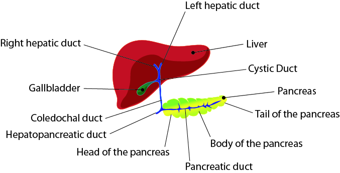
  • Nose: The nose has an external and a internal part. The external part is divided into two nasal channels called nostrils. The inner part is a large cavity located between the facial bones, just above the mouth. The floor of the nasal cavity is the hard palate, and the roof is a part of the ethmoid bone called cribriform plate. It is divided into two parts, the right and the left ones. It is connected to the pharynx through two openings called choanes. The nasal cavity is responsible for filtrating, heating and wetting the inhaled air. It also receives olfactory stimulus and modifies our voice.
  • Pharynx: This is a thirteen centimetre long duct similar to a funnel, that connects the nasal cavity with the cricoid cartilage, that it is the upper part of the larynx. It has three parts. The upper part is located just beneath the nasal cavity and is called nasopharynx. The nasopharynx is connected with the nasal cavity through the choanes, and with the mid ear through the Eustachian tube. The next part of the pharynx is the oropharynx, and it is located behind the oral cavity. The oropharynx connects the mouth with the respiratory and the digestive systems. The lower part of the pharynx is the hypopharynx, that connects the oropharynx with the oesophagus (digestive system) and the larynx (respiratory system).
  • Larynx: It is a short duct, made up of nine cartilaginous pieces, that connects the pharynx with the trachea (windpipe). The larger piece of the larynx is the thyroid cartilage, that it is also known as the Adam’s apple. This structure covers the thyroid gland. Another important piece is the epiglottis, a sort of flap that opens and closes the duct in order to prevent food from going into the trachea. The vocal folds are also located in the larynx.
  • Trachea: This is a twelve centimetre long and two and a half centimetre diameter duct that connects the larynx with the bronchi. It lies just in front of the oesophagus. The trachea is surrounded by fifteen incomplete cartilaginous rings, that protect and maintain the airway. This rings are C shaped, to allow the dilation of the oesophagus when the food is passing through it. At the level of the fifth dorsal vertebra the trachea bifurcates into two principal bronchi.
  • Bronchi: The trachea bifurcates into two principal bronchi, called right bronchus, that enters in the right lung and left bronchus, that enters in the left lung. After penetrating in the lung, each bronchus divides into two secondary bronchi. The secondary bronchi divides into tertiary bronchi. Each tertiary bronchi divides into two bronchioles. The bronchioles continue dividing successively, completing sixteen total divisions. The air that fills the bronchi and bronchioles (around 150ml) is not used to breathe, because there are not special structures to allow the exchange of gases, so it does not takes place. The structure where this exchange of gases takes place is called alveoli and these are located after the last bronchiolar division.
  • Lungs: the lungs are two large conic shaped organs, located in the thoracic cavity and separated by the heart and the mediastinum. Each lung is covered by two membranes. The outer one is attached to the thoracic wall and it is called parietal pleura. The inner one is attached to the lung surface and it is called visceral pleura. Between both layers there is an internal fluid that keeps both membranes together and that lubricates them the to avoid friction when they move. This liquid is called pleural effusion. The lower and broader part of the lung is called base of the lung. The upper part of the lung is called anterior border. The cavity from where blood vessels and bronchi enter is called hilum. The right lung is slightly larger than left one, because the left one must leave a space for the heart. The left one is, however, longer than right one, because it must leave a space for the liver. Both lungs have fissures that divide them into lobes. The left lung has one fissure that divides it into an upper and a lower lobe. The right lung has two fissures that  divide it into a lower, a middle and a upper lobe. The lungs are the organs where the bronchioles divide. After the last division, the alveolar sacs can be found. Each one of these sacs has two or three alveoli. The alveoli are covered by blood vessels, because these are the anatomical structures where the exchange of gases between the air and the blood takes place.
Respiratory System: Anatomy.

Physiology of Respiration.
Introduction.
First, we will study the process called pulmonary ventilation, that explains how the air flows from the exterior of our body to the lungs and from the lungs to the exterior of our body. Then, we will study the exchange process between the air and the blood, that takes place in the pulmonary alveoli and it is called external respiration. Finally we will study the exchange of gases between the blood and the internal tissues, that is called internal or tissue respiration.
Pulmonary Ventilation.
Pulmonary ventilation, also called breathing, is the movement of the air between the exterior of the body and the lungs. The air enters the lungs from the environment to in a process called inhalation. The air exits the lungs to the environment in a process called exhalation. These movements of air are a consequence of the changes of pressure in the lung and of the special properties of this organ, that is capable of increasing its volume by distension and to return to its original size by elasticity.
The process of entrance of air into the lungs is called inhalation. It takes place when the lungs expands, increasing their volume. The expansion results from the contraction of the respiratory muscles: the diaphragm and the internal intercostal muscles. The most important muscle is, by far, the diaphragm. When it contracts, its convex morphology changes, becoming flatter. This movement pulls the lung down, enlarging its lower part. The internal intercostal muscles raise the thoracic cage, causing the expansion of the lungs because they are closely attached to the ribs. These two processes increase the volume of the lungs. The higher volume leads to a drop in the internal pressure, so that the air moves from the environment to the lung.
The release of air is called exhalation. It is a passive process, no muscular contraction is required. The elastic fibres of the lungs and the weight of the thoracic cage decrease the volume of the lungs when the respiratory muscles relax. The reduction of volume leads to an increment of the internal pressure, so that the air moves from the interior to the exterior.
Although this is a passive process, the contraction external intercostal and the abdominal muscles can accelerate the release of air. This is called forced exhalation, and is carried out when the body needs to improve the exchange of air.
Ventilation Volumes.
During normal breathing around 500ml of air is exchanged between the lungs and the environment. This amount of gas that enters and afterwards exits from the lungs is called Tidal Volume (VT).
Not all this gas is available to exchange oxygen and carbon dioxide. Around 150ml of air never reaches the alveoli and stand in the outer respiratory ducts: nasal cavity, pharynx, larynx, trachea, bronchi and bronchioles. This volume that is not directly used in the pulmonary respiration is called Dead Space (DS).
The Respiratory Minute Volume (MV) is the amount of air exchanged between the lungs and the environment per minute. An adult human being breathes approximately  twelve times per minute, exchanging 500ml per breathing (Tidal Volume), so it is easy to calculate that the MV is 6000ml/min. 
We can breathe more deeply, inhaling more than 500ml. We can reach between 3000ml and 3500ml more in a forced inhalation. This is called Inspiratory Reserve Volume (IRV). We can even take more air if, just before the forced inhalation, we exhale as much air as we can. This air that we can release through forced exhalation, around 1200ml, is called Expiratory Reserve Volume (ERV).
After exhaling all the air that forced expiration allows, there is a volume of air that remains in the respiratory system. This amount of gas that we cannot release is very important, because it prevents the duct and alveolar sacs from collapsing. It is 1200ml more or less, and it is called Residual Volume (RV).
If we add the Tidal Volume to the Inspiratory Reserve Volume we obtain the Inspiratory Capacity (IC). It is around 3600ml. If we add the Residual Volume to the Expiratory  Reserve Volume we obtain the Functional Residual Capacity (FRC). It is around 2400ml.
The Inspiratory Reserve Volume added to the Tidal Volume and to the Inspiratory Reserve Volume is called Vital Capacity. It is around 4800ml. If we add all the volumes (IRV+VT+ERV+RV) we obtain the Total Lung Capacity (TLC). It is around 6000ml. 
Ventilation Volumes

Pulmonary Respiration Physiology.
The physiology of pulmonary respiration is based on the concentration gradients or differences in partial pressure. The internal membrane of the lungs is extremely thin (around 10.5μm), so that the gases are easily exchanged. And the internal surface of the lung is really broad, around 70m2 if we count all the alveolar surface.
The air that reaches the alveoli is very rich in oxygen, between 100-105mmHg. The concentration of oxygen in the blood in the capillaries of the lung, however, is quite low, around 40mmHg. Due to this, the oxygen tends to flow from the air to the blood, until both concentrations become equivalent. When the blood exits the capillaries of the lung, its concentration of oxygen is approximately 110mmHg.
To improve the movement of oxygen in the blood, it is transported linked to a special protein called hemoglobin.
Carbon dioxide concentration in the air is around 40mmHg. When the blood arrives in the alveoli, its concentration of carbon dioxide is around 45mmHg. Due to this, the carbon dioxide tends to flow from the blood to the air until both concentrations become equivalent. When the blood exits the capillaries of the alveoli the concentration of carbon dioxide is 40mmHg.
The carbon dioxide is not transported by any protein, but is transformed into a different substance called bicarbonate.
Tissue Respiration Physiology.
The situation in the tissues is just the opposite to in the lungs. The extracellular fluid that surrounds the cells is very poor in oxygen, because it has been consumed by the cells. Its concentration is 40mmHg. As we studied, the concentration of oxygen in the blood that comes from the lungs is 100mmHg. Due to this, the oxygen tends to flow from the blood to the tissues until both concentrations become equivalent.
Carbon dioxide, however, is more concentrated in the extracellular matrix, because the cells produce and release this substance during their metabolic activity. Its concentration is 45mmHg, whereas in the blood the concentration is around 40mmHg. So the carbon dioxide flows from the extracellular matrix to the blood until both concentrations become equivalent.
A part of this carbon dioxide is transported by the blood linked to hemoglobin, but only a low quantity, around the 23%. A small amount, the 7%, is transported dissolved in the plasma. The rest of the carbon dioxide, around the 70%, is transformed into bicarbonate by an enzyme called carbonic anhydrase. It is so how it is transported, because this substance can be easily dissolved in the plasma.
Respiratory System: Anatomy.

Control of Breathing. 
Breathing is an extremely controlled process, because it must be finely adjusted to the requirements of the body. An ordinary human being consumes around 200ml of oxygen per minute. While intense exercising, however 30 times this amount can be consumed. To increase the amount of taken oxygen the body increases the respiratory rate and depth.
The respiratory rate at rest is controlled by some areas of the nervous system located in the bulb and the pons. The Bulb Rhythmic Area controls the basic system of respiration and the respiratory rate at rest. The Pneomotaxis Centre controls the coordination between inhalation and exhalation. The Apneustic Centre controls the inhalation.
Other zones of the brain have connections to these respiratory centres and they can raise or decrease the respiratory rate when it is necessary. When the pH of the blood decreases, for instance, it is related to an increment of the bicarbonate dissolved in the plasma, so the respiratory rate must be increased to release the excess of carbon dioxide. When some receptors detect that the amount of oxygen drops, they promote an increment of the respiratory rate. There are many different chemical receptors in out body, such us the carotid and aortic receptors.

Some hormones can also have different effects on the respiratory system. Adrenalin, for instance, affects not only to the respiratory rate, but also to the amount of air inhaled by changing the diameter of the bronchioles and increasing the air flow to the alveoli. Other hormones have just the opposite effects.

domingo, 24 de enero de 2016

Digestive System

Digestive System.
Kinder beim Mittagessen, M.L.Benjamin Vautier
The digestive system is the main one responsible for processing the food we consume, so that it can be used as an energetic support or as a font of source materials.
The digestive system fragments large molecules of the food transforming them into smaller molecules that can be absorbed. In this unit we will analyse the anatomy, physiology and reproduction of eukaryotic animal cells.
The whole digestive process can be divided into five consecutive actions.
  • Ingestion: the food passes from the exterior to the interior of the body via the mouth.
  • Movement of the food: the food moves across the digestive tube.
  • Digestion: fragmentation of the food by mechanical and chemical processes.
  • Absorption: the nutrients obtained when the food is digested are transported from the gastrointestinal duct to the blood or lymphatic system.
  • Defecation: release of non-digested substances.
Anatomy of the Digestive System.
Introduction.
The digestive system can be divided into two parts:
  • Gastrointestinal duct: it is a nine meter long tube that starts in the mouth, crosses the thorax and abdomen and ends at the anus.
  • Accessory structures: there are several different structures. The most frequent ones are glands, although there are other structures such as teeth or the tongue.
In this unit we will study the digestive duct, from the mouth to the anus, analysing the most relevant accessory structures in each part.
Mouth.
Also called oral cavity, this is the gate where the ingestion of food takes place. It is bound laterally by the cheeks, at the right and left sides, the hard and soft palates at top and the tongue at floor. It is separated from the exterior by two fleshy folders covered by thin skin called lips.
The inner part, between the cheeks is called vestibule. Behind the vestibule, the duct that binds the mouth posteriorly is called isthmus of the fauces. This duct separates the mouth from the pharynx. Just in the boundary of the isthmus there is a little hanging piece of soft tissue called uvula.
The roof of the mouth is called the palate and has two parts. The anterior part is made of bone and it is called hard palate. The bones that form the hard palate are two small palatine bones and a part of  the maxillary bone. The posterior part is made of muscle and it is called soft palate.
There are also several accessory structures in the mouth:
Tooth Anatomy
  • Tongue: this is a skeletal muscle that forms the dynamic structure of the mouth floor. It is essential to break the food down using the teeth and then to swallow the bolus. The taste sensor is also located on the surface.
  • Salivary Glands: they constantly secrete saliva to the mouth. So that they wet the oral cavity and the pharynx. When the food enters into the mouth the secretion rises. Saliva lubricates the food and dissolves part of its components, starting the chemical  digestion of several components. There are three pairs of salivary glands. One pair are called Parotids and they are located beneath the ears. Another pair of glands are called Submandibular and they are located in the posterior part of the mouth floor, just beneath the tongue base. The last pair of glands are called Sublingual and they are located in the anterior part of the mouth floor, under the central part of the mouth.
  • Teeth: accessory structures located in the alveolar apophysis of the maxillary bones and mandibular bone, which are covered by the gums. They are very hard and resistant. When the food is chewed, the teeth cut and crush the food. Adult human beings have 32 teeth. There are four types of teeth and adult human beings have 8 incisors, 4 canines, 8 premolars and 12 molars. 
Mouth: Teeth.

Pharynx.
This duct is a part of the digestive and respiratory systems at the same time. The contraction of the muscles that surrounds the tube moves the food from the mouth to the oesophagus. It is in contact with upper part of the larynx, called epiglottis, that prevents the food from passing into the respiratory system.
Oesophagus.
This is a 20 to 30 centimetre long tube, located beneath the windpipe (trachea). It starts at the lower portion of the pharynx, crosses the mediastinum trespassing the diaphragm through the oesophagic hiatus and ends up at the upper portion of the stomach. 
The peristaltic movements of the wall transport the bolus from the pharynx to the stomach. The tube terminates at the cardias, also called superior oesophageal sphincter, that controls the entrance to the stomach.
Stomach.
Dilated portion of the gastrointestinal duct, located under the diaphragm. Its shape and size changes according to the movements of the diaphragm, the amount of food and the digestion phase. It has four parts. The upper part, called cardia, has the opening to the oesophagus. Just under the cardias, at the left side, there is a large concave part called fundus. The central part of the stomach is called body. And the lower part, connected to the intestine by a sphincter called pylori, is called pyloric antrum.
Stomach Antomy.
Cardia and pylori are two sphincters stat enclose the food in the stomach during the digestive process of the stomach, so that the food being digested and the digestive acids can not flow back to the oesophagus or pass into the intestine until the process has finished.
Two important digestive processes take place in the stomach: mechanical digestion and chemical digestion. The mechanical digestion is carried out due to the peristaltic movements of the stomach wall. These movements also mix the food with the gastric juices, released by the stomach wall. These juices are responsible for the chemical digestion of the food. The main component of the liquid that carries out the chemical degradation of food components is the hydrochloride acid (HCl), although there are other digestive components, such us enzymes responsible for degradation of proteins, fats or polysaccharids. The bolus after being digested in the stomach is called chyme. 

Small Intestine.
It is a six meter long and three centimetre thick tube, that stats at the pyloric sphincter, just beneath the stomach and ends up at the ileococcal sphincter, that separates the small intestine from the large intestine. It is intricately tingled in the central and inferior region of the abdominal cavity.
The small intestine has three parts. The first one is called duodenum, it is only 25 centimetre long and it is located just beneath the stomach. It is an important region, because liver and pancreas release their secretion to it. The central part of the small intestine is called jejune and it is 2.5 meter long. The final part is between 3.5 and 4 meter long and it is called ileum. The ileocecal sphincter separates the ileum from the large intestine.
The small intestine wall is not smooth, but it is profusely folded, forming lots of little finger shaped protrusions called intestinal villi. Their function is increasing the intestinal surface, so  intestine has an extremely huge area from which to absorb nutrients from the food. The epithelial cells that cover the inner part of the tube, besides, have microvilli in their upper membrane, to increase the absorptive surface even more.
As we have mentioned, the absorption of nutrients takes place in this part of the digestive system. In fact, it is the only place where a massive absorption takes place. And it is also the place where the digestion of food is completed, although it has started in the mouth and above all in the stomach. The mechanical digestion in the small intestine, led from the peristaltic movements of the wall, is not really relevant. The chemical digestion, however, is very important. It transforms the chyme from the stomach into chyle. The cells of the intestinal wall release little amounts of digestive products, although chemical digestion is principally carried out by chemical substances released to the intestine by two accessory glands: pancreas and liver.
  • Pancreas: this is a 12.5 centimetre long and 2.5 centimetre thick (although the thicker part is usually slightly thicker) gland. It is located beneath the major curvature of the stomach. It is connected to the small intestine by two ducts. The gland has two parts, the thicker part near duodenum and called the head, and the thinner part, slightly sharp called the tail. The pancreas has two main functions. On the one hand, pancreas produces and releases pancreatic juice, a liquid rich in enzymes responsible for degradation of nutrients: amylases that degrade sugars, proteases that degrade proteins and lipases that degrade lipids. On the other hand, it produces and releases digestive hormones that control the levels of glucose in the blood. This function is carried out by a section of the pancreas called endocrine pancreas. The pancreatic duct that connects the pancreas with the intestine joins with the bile duct from the gallbladder.
  • Liver: this is the largest gland of our body. It weighs around 1.5 kilogrames. It is located under the diaphragm, taking up a big space of the right hypocardiac and a part of the abdominal epigastric. It is divided into two big lobules, called left and right lobule, joined together by the falciform ligament. It is irrigated by the hepatic arteries, that provide oxygenated blood, and by the inferior vena cava, that arrives from the small intestine and provides nutrients. The liver produces and releases between 800 and 1000ml of gall per day. This pale green liquid is responsible for increasing the pH of the food that has been digested n the stomach, and for promoting the emulsion of lipids and fats, so that large a cumulus of fats are transformed into little drops of lipids that can be easily absorbed. The gall is also used to release waste products produced by the liver, such as bilirubin, made from destroyed erythrocytes and responsible for the brown colour of the faeces. Gall is produced by the liver, but it is not directly released to the intestine, but is stored in the gallbladder. 
  • Gallbladder: it is a pear-shaped sac, where gall is stored. It is located just under the lower depression of the liver. The bile duct connects the gallbladder with the pancreatic duct, and both ducts joined connect with the intestine.
Anatomy of liver, gallbladder and pancreas.

Large Intestine.
This is a 1.5 meter long and 6 meter thick tube, that starts at the cecum and ends up in the anus. Large and small intestine are separated by the ileocecal sphincter. There is a 6cm long section of tube just below the sphincter, called cecum. At the end of the cecum a little cylindrical structure called vermiform appendix can be found. The appendix and cecum are the right side of the abdomen. The rest of the large intestine is called colon and can be divided into several parts. First, the ascending colon from the ileocecal sphincter to the right-upper part of the abdomen, just beneath the diaphragm. Then the colon turns 90° in the right colical angle. The portion of colon that crosses the abdomen from the right to the left side of the abdomen, parallel to the diaphragm axis, is called transverse colon. It turns 90° again in the left colical angle, and crosses the abdomen from the left-upper to the left-lower part of the abdomen. It is called descendant colon. The descendant colon turns 90° again just when it is parallel to the iliac crest, and then is called sigmoid colon. The tube turns again and this final part is called rectum. In the last 3cm of the rectum there is a large sphincter called anus, that controls the release o faeces to the exterior. The colon is the place where non digested and non absorbed substances arrive. There are many bacteria that transform these into waste product. Some vitamins are produced by these bacteria, and can be absorbed by large intestine. Waste products, however, are not digested or absorbed, only a large amount of water from the faeces is absorbed, to prevent our body from dehydration. So that the faeces produced after a correct digestive process are nearly dry, and made up of substances that our body can not digest or absorb. 
Digestive System.
Physiology of the digestive system.
The whole digestive process is based on the physical and chemical degradation of the food in order to transform the complex molecules into small and simple ones that could be absorbed and transported from the interior of the small intestine to the blood or the lymph. Non digested or absorbed substances must be dried, condensed and released to the exterior.
This digestive process starts in the mouth. Teeth cut the food in small pieces and some chemical components of saliva carry out the first chemical digestive step. Salivary amylase degrades complex sugars, for instance, transforming them into smaller and simpler sugars. The food, after being chewed and mixed with saliva is called bolus.
After being swallowed the bolus descends along the oesophagus, arriving at the stomach. The bolus is enclosed in the stomach, because two sphincters, cardiac and pylori, are closed. So that the bolus cannot pass to the intestine until it has been digested, but cannot either flow back to the oesophagus.
The stomach wall releases extremely acidic gastric juices, rich in degradative enzymes that destroy large molecules, transforming them into smaller ones, such as the enzyme called Pepsine, that breaks down proteins transforming them into simple amino acids. The stomach wall, besides, carries out abrupt peristaltic movements that provoke the mechanical digestion of food and improve the chemical digestion mixing the food with the gastric juice. The food after being digested by the stomach is called chyme.
The final chemical and physical digestive process of the food takes place in the small intestine. The chemical digestion in the small intestine is carried out by the pancreatic juice. This liquid is rich in degradative enzymes: proteases that destroy proteins transforming them into amino acids, lipases that destroy fats transforming them into simple lipids and amilases that destroy polysaccharides transforming them into simple sugars. Pancreatic juice and gall also neutralise the pH of the chyme, which is very acid due to the hydrochloride acid of the stomach. Emulsification of fats is another important function of gall. This emulsification is essential to improve the absorption of lipids and fats by the small intestine.
The food after being digested in the small intestine is called chyle. And chyle is the final product ready to be absorbed by the small intestine. As we studied before, the inner wall of the intestine is profusely folded to increase the surface in contact with the food, improving the absorption of nutrients. The exterior of the small intestine is highly vascularised, so that the nutrients are transported directly from the inner part of the intestine to the blood vessels and lymphatic system. The transportation of nutrients is a controlled process and only small digested substances, such as simple sugars, amino acids or small fats are able to trespass the barrier. The fats are mainly transported by the lymphatic system, whereas the rest of  nutrients are transported by the circulatory system.
Large intestine hardly absorbs any nutrient, only some vitamins released by bacteria and, above all, large amounts of water. The inner surface of the large intestine is smooth, with no folds (merely because they are not necessary). The absorption of water is extremely important, preventing our body from dehydration.
After this process, all the dehydrated remains, made up of non digested and non absorbed substances reach the end of the tube and are released, during the defecation, through the anus.
As we studied, liver is responsible for provision of gall, but it has also other important digestive functions. First, it is responsible for storing some nutrients, such us sugars (forming a macromolecule made of large chains of sugar called glycogen) and fats. Second, liver transforms toxic products, such as cholesterol and bilirubin and releases them into the intestine, so that they are expelled them to the exterior. And third, liver is responsible for many metabolic reactions, such us gluconeogenesis, a group of chemical reactions essential to produce glucose from other simpler metabolic products.
The pancreas is very important to control some digestive processes. It produces two hormones, insulin and glucagon, that control the glucose balance. Insulin is produced when the glucose level rises, promoting actions to reduce it. On the other hand glucagon is produced when the glucose level drops, promoting actions to increase it. This actions are very important, because the glucose level in blood must stay relatively constant.
Digestive System


domingo, 27 de septiembre de 2015

Health and Illness

Health and Illness: Definition.

The most simple definition for health is the absence of illness. In other words, we can say that we are healthy when we are not ill. But, when are we ill? When are we healthy? What is exactly a disease?
The World Health Organization defines health as the state of complete physical, mental and social well-being and not merely the absence of disease or infirmity.

In this definition, health is divided into three basic parts: physical, mental and social. Physical health, also called allostasis, is the maintenance of physiological homeostasis through changing circumstances. Mental health is related to the sense of coherence. And finally social health is the ability to manage life and to participate in social activities.

On the other hand, we define illness as an abnormal or pathological condition that affects part or all of one organism. Diseases are disorders in system, organs, tissues or any other structure of the body.

Types of Diseases.

Diseases can be divided into two different groups: communicable diseases and non-communicable diseases.

Communicable diseases are also called infections. They are related to the invasion of our body by infective agents, such as viruses and bacteria.

Non-communicable diseases, however, are not related to infections. They are defined as diseases or medical conditions which are non infectious and non transmissible among people.

Summing up, ordinary diseases transmitted among people, such us flu, AIDS, or salmonellosis are communicable diseases. Injuries due to accidents, congenital or hereditary affections are non-communicable diseases.

Communicable Diseases.

As we studied, communicable diseases are related to the invasion by infectious agents. These agents are also called pathogens, and can be defined as living beings with the ability to cause disorders in their hosts.

The pathogens can be divided into two groups: microscopic and macroscopic parasites. The microscopic parasites cannot be seen withe the naked eye, whereas macroscopic parasites can be seen with the naked eye or a magnifying glass.

The most important microscopic parasites are viruses, bacteria, protozoa, fungus and prions. The most important macroscopic parasites are nematodes, insects and arachnids.

Viruses.

Viruses are extremely tiny and simple organisms, so small that they only can be seen using an electron microscope.

Viruses are made up of three main components: a nucleic acid, proteins and and in some viruses an envelope of lipids.

The nucleic acid is the most important part and its function is storing the information. The nucleic acid can be DNA or RNA, and encodes information about how to produce the rest components of the virus and how to reproduce. The life cycle of the virus consists of invading a cell and using this cell to produce multiple copies of its nucleic acid.

Viruses have two kinds of proteins: structural proteins and enzymes. The structural proteins make up the capsid of the virus. The capsid is the protective coat of the organism where the nucleic acid is enclosed. The enzymes are proteins that carry out the chemical activities of the virus. These activities are, mainly, duplication of DNA and production of the proteins of the new viruses.

VIH diagram
The envelope of lipids appears in some viruses, coming from the host cells. So the envelope is not produced by the virus, but is stolen from the host cell.

There are many examples of diseases caused by viruses. The most important ones: flu, AIDS, rabies, poliomyelitis, rhinitis, ebola, hepatitis, herpes or measles.

Bacteria.

Bacteria are simple living beings that belong to the kingdom Monera. They are prokaryotic cells, so they are always unicellular organisms. They are simple cells with neither nucleus nor internal complex organelles.

This simple cells are covered by a proteinic cell wall. They reproduce by partition. And most of them are not pathogen. In fact, many of them are useful; they are related to the production of some food, such as yogurt for instance. Besides many bacteria live in our body but do not cause diseases. Some of them are essential in our body, releasing proteins during our digestion or preventing our body to be invaded by other bacteria.

Pathogenic bacteria can produce toxins and attack our cells. Some bacteria that are not dangerous in one part of the body, can cause grave diseases in another. There are bacteria, for example, that live in our skin and do not cause any kind of disease, but can be extremely dangerous if they reach internal organs traveling through the blood.


There are many examples of diseases caused by bacteria. The most important ones are typhus, salmonellosis, cholera, poliomyelitis, gastroenteritis, plague, syphilis, tetanus, pneumonia or anthrax. 
   
Protozoa.

Protozoa are complex cells that belong to the kingdom Protoctist. So they are eukaryotic unicellular organisms. They are cells with nucleus and internal organelles, covered by a plasmatic membrane but not by cell wall.

Just like bacteria, they reproduce by partition.

Most of them are not pathogen. There are many protozoa mainly living on organic matter, because they are not autotrophs, so they can not produce organic matter from inorganic matter and must take the organic matter from their environment. Due to this, the growth of protozoa is usually associated with bad hygienic conditions and places where organic matter is specially abundant.

Pathogenic protozoa can produce toxins and attack our cells. There are not many diseases related to protozoa, although one of them, called Malaria, is a extremely grave disease that causes thousands of deaths year after year, mainly in tropical regions of Africa.

Toxoplasmosis, trypanosomiasis or leishmaniasis are other relevant diseases caused by protozoa.

Fungi.

Fungi belong to the Kingdom Fungi. They are unicellular or multicellular organisms, made up of one or more eukaryotic cells. So that these cells have nucleus and internal complex organisms. They are covered by a plasmatic membrane, but not by cell wall.


Multicellular fungi form structures called hyphae and they usually divide by simple partition, although they also have sexual reproduction.

Most of them are not pathogen and there are many fungi that are commonly used in industrial processes, such as production of cheese or fermentation of fruit to obtain alcoholic drinks: cider, wine, beer, whiskey, etc.

The most frequent diseases produced by fungi are superficial affecting our skin. Internal infections by fungus are not usual. Tinea pedis (better known as ringworm), candidiasis or pityriasis are examples of diseases caused by fungus.

Prions.

Prions are not living beings, they are simple proteins, similar to toxins, that some animal tissues can build up. They can enter in our body across the digestive or respiratory system, reaching our cells and provoking formation of irregular proteins.

There are few diseases known, but one of them has been sadly famous throughout the last years. It affects cows and sheep, it is called bovine spongiform encephalopathy, or mad cow disease. When it is transmitted to humans (due to the consumption of infected food), the prion causes a disease called Creutzfeldt-Jakob disease.

Epidemics and Pandemics.

When a disease spreads affecting many people, we can talk about epidemics and pandemics.

Epidemic is defined as a widespread disease that affects many individuals in a population at the same time period.

Pandemic is defined as an epidemic disease that spread through human population across a large region, frequently multiple countries or even continents.


Macroscopic parasites.

These invaders are visible with the naked eye or using magnifying glasses. Many of them can not only cause direct diseases, but also transmit other diseases. Then, the parasite is called vector.
So vector is defined as any agent that carries and transmits an infections pathogen into another living being. Probably the most famous vector is the mosquito that transmits malaria.

Most of the macroscopic parasites feed from the host organism. Lice, for instance, live in our hair and feed from our blood. Parasites are usually very specialised, so that a parasite can live only in one or a few species of host.


Macroscopic parasites can be classified according to the animal group they belong to. Human beings can be infested by nematodes, such us roundworms that invade our intestine. Or Platyhelminthes, such as taenia, also called genus, a flat worm that invades our intestine.

The most abundant macroscopic parasites are, by far, arthropods. There are many insects that parasite human beings, such as fleas, mosquitos or lice. But there are also arachnids, such us ticks and scabies.  
  

Non communicable diseases.

Non-communicable diseases are non infectious diseases, so that they are not passed from person to person. According to their origin or characteristics, there are many types of noncommunicable diseases.

Congenital diseases are diseases that exists at birth. Spina bifida, for instance, that is characterised by the incomplete closing of the backbone during the embryonic development (it is related to deficiency of folic acid during pregnancy).

Hereditary diseases are transmitted from parents to descendants. Cystic fibrosis, for instance, is a severe disorder that affects the lungs (and also other organs, such as liver or pancreas).

Chronic diseases are prolonged in time with no possible cure. Many cardiovascular diseases evolve in chronic diseases.

Deficits are diseases related to low amounts or lacking in any important substance. Vitamin C lacking or deficiency, for instance, results on a disease called scurvy.

Genetic diseases are related to dysfunction in genetic information. Down syndrome, for example, is a disorder caused by the presence of an extra copy of the chromosome 21.

Traumatic diseases result from accidents or injuries. Broken bones, burns or bruises are typical examples.

Degenerative diseases are progressive disorders in some organs or structures. Parkinson’s disease or Alzheimer’s disease are examples of degenerative diseases that affect the central nervous system.

Functional diseases are diseases in tissues, organs or systems for different reasons. Diabetes, for instance, is a disease related to pancreatic disfunction.

Fighting against illness.

There are two important concepts when we talk about fighting against illness: prevention and cure.
Prevention could be defined as the actions carried out to preserve the health. Curation, on the other hand, could be defined as the actions carried out to recover the health. Obviously, preventing is more important because it is better to preserve our body from illness than to recover our health after getting ill.

Prevention.

Healthy habits are, by far, the most important way to prevent diseases. There are many habits that help us to preserve our body from ordinary diseases.

Doing regular physical exercise, for instance, is very useful to prevent heart and circulatory diseases, obesity, stress and other psychological alterations.

A balanced or healthy diet is important to prevent obesity or anorexia. It helps us, besides, to ensure the consumption of nutrients in optimum amounts.

Improving hygienic conditions is essential to prevent infections. Brushing our teeth to prevent cavities or washing our hands to prevent infections are two typical examples.


Having a healthy lifestyle is also very important. Consuming drugs or other toxic substances, such us alcohol, clearly increases the probability of suffering from severe diseases, affecting diverse vital organs and reducing our life expectancy.

Our lifestyle is also related to the prevention of accidents. Driving carefully and respecting safety rules when we work with dangerous devices or machines help us avoid unexpected and serious accidents.    

Preventive medicines: Vaccines.

Vaccination is defined as the administration of substances to stimulate the adaptive immune system.

The inoculated substance could be made up of parts of microorganisms (viruses and bacteria) or attenuated pathogens (in other words, pathogens unable to provoke diseases). When the immune system finds these substances it produces antibodies. Antibodies are complex proteins that destroy the invasive agents. Therefore, if a real infectious agent  invades our body, our immune system has already produced antibodies, so the response will be faster and more effective.

Summing up, vaccines are very important because they teach our immune system how to fight against dangerous microorganisms.

Vaccination has been transcendental in defeating some serious diseases, such as smallpox or poliomyelitis. In developed countries there is an official vaccination timetable, so that some severe infections, such us rabies, have nearly disappeared in these regions.

Vaccination is extremely useful to prevent infection, but it is not the best system when the infection has already taken hold.

Curation.

When we are sick we use medicines to recover our lost health. There are two main groups of medicines: antibiotics and other generic medicines.

Antibiotics.

Antibiotics are substances that kill or inhibit the growth of microorganisms. So that they must be used to fight infectious diseases.

Most antibiotics are specific to a few different organisms. Due to this, knowing the microorganism responsible for the infection is quite important to select the best antibiotic to  fight against it.

Antibiotics are toxic substances, so they must be administrated carefully. When microorganisms are attacked by antibiotics, they try to create defences against that substance. For this reason, an inappropriate use of antibiotics can create resistant microorganisms. These resistant microorganisms are quite dangerous, because when they cause infections the classical antibiotics are not effective, so destroying them could be extremely difficult.

Viruses do not have their own metabolism, they use our cells to reproduce. Due to this, antibiotics are not effective when we want to fight viruses.

Other medicines.

There are other types of medicines, useful when the illnesses are not related to infections or when we want to mitigate the undesirable effects of any affection.


Analgesics, also called painkillers, are medicines used to relieve pain. They are used, for instance, when somebody has a headache or a toothache. Antipyretics are substances that reduce high temperatures. High temperatures are frequently associated with infections.  Anti-inflammatories reduce inflammation. Inflammation is a response by our body to infections or other injuries. Bronchodilators dilate bronchi. Antihistaminic are used to fight allergic reactions. There are many others medicines, such as laxatives that increase the intestinal movements when you are constipated, vasoconstrictors and vasodilators to control the blood pressure, stimulators, anti-stress, mood stabilizers and tranquilizers to regulate neural activity, antitussives to reduce the intensity of coughs, etc.